BoneMRI SIJ
Main Investigator: Lennart Jans
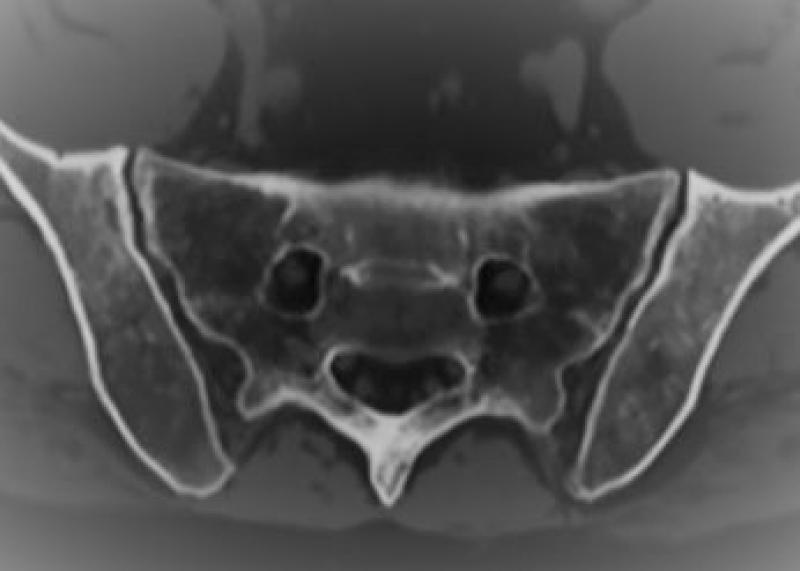
BoneMRI allows to generate 'CT-like' images from MR images applying an AI trained model. In rheumatic disease, this allows us to depict structural lesions such as erosions and new bone formation without radiation.
Normal variance SI Joints in children
Main Investigator: Nele Herregods
The normal appearance of the growing bone may mimick inflammatory disease and, moreover, normal variants may occur. We seek to find MRI features that differentiate normal growth from disease.
DECT
Main Investigator: Lennart Jans
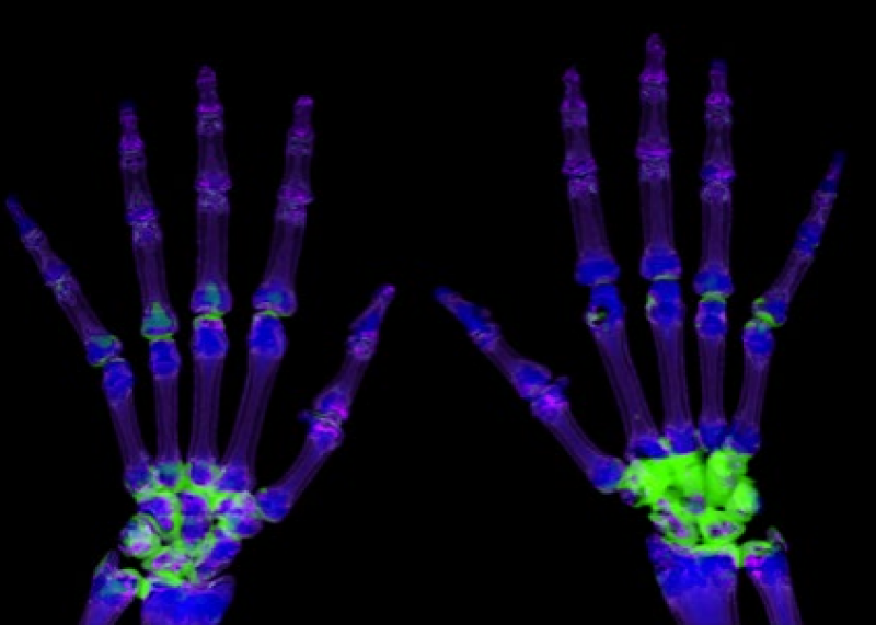
Dual Energy CT is a new technique that enables to depict inflammatory bone marrow oedema in patients with rheumatic disease. In patients with contraindications to MRI, DECT can thus detect active inflammation.Dual Energy CT is a new technique that enables to depict inflammatory bone marrow oedema in patients with rheumatic disease. In patients with contraindications to MRI, DECT can thus detect active inflammation.
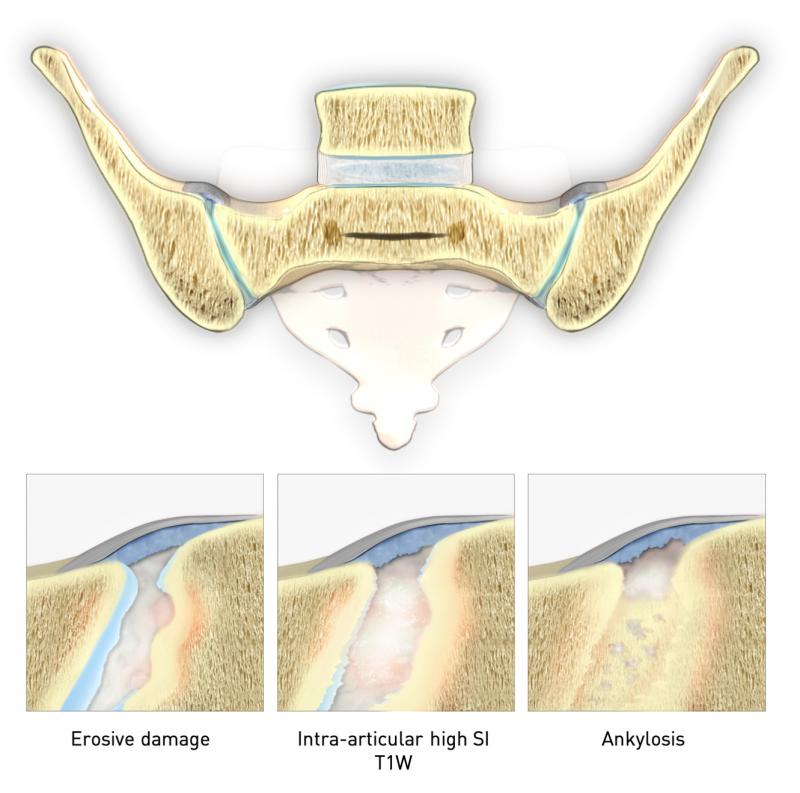
New bone formation on MR of the sacroiliac joints and the spine in spondyloarthritis
Main Investigator: Frederiek Laloo
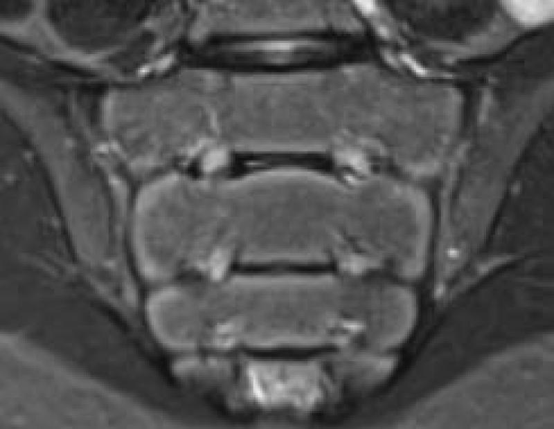
Spondyloarthritis has two hallmark features: active inflammation and structural lesions with new bone formation. There is increasing interest in the role of structural lesions at MRI, as various established features of new bone formation can be evaluated at MRI and new features such as ‘backfill’ can even help make a diagnosis. Our past research has helped determine the diagnostic value of these features at MRI, and future research will focus beyond the scope of diagnostic value, e.g. disease progression.
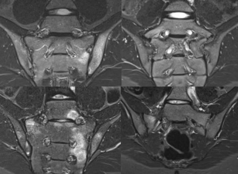
DD of BME
Main investigator: Eva Schiettecatte
Not all bone marrow edema is inflammatory. Trauma, tumor, infection, normal variants can lead to BME just as well. We study the prevalence of BME of the SI joints and spine, and describe the typical imaging features of BME related to its etiology.
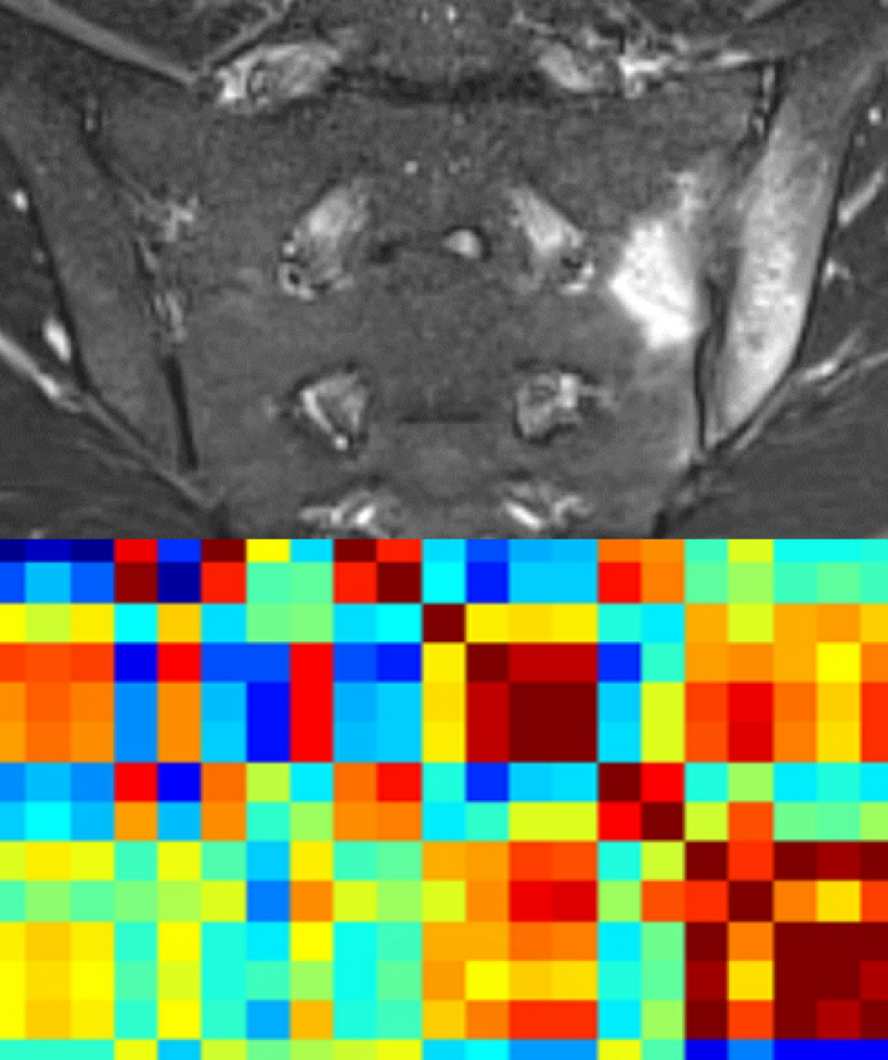
Analysis with AI in spondylarthropaties
Main Investigator: Thomas Van Den Berghe
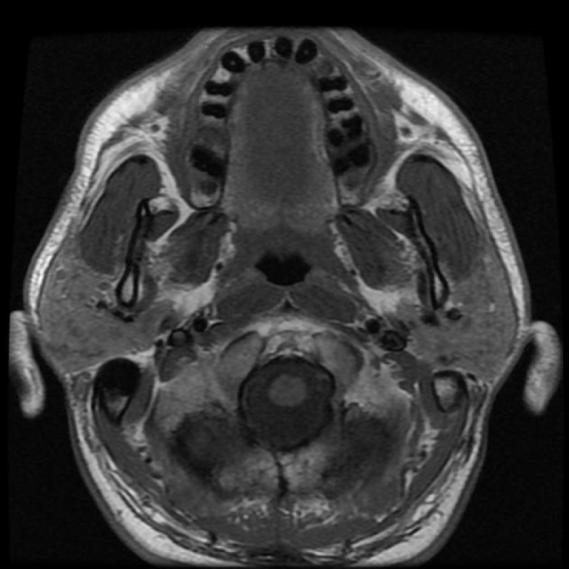
Radiomics, radiogenomics and analysis of medical imaging techniques with artificial intelligence in patients with plasma cell dyscrasias and spondylarthropathies. Quantitative evaluation of disease status on anatomical, DCE and DWI MRI sequences.
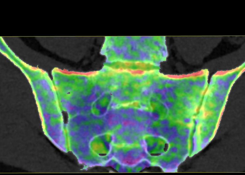
Application of dual-energy CT for bone marrow edema detection in the sacroiliac joint.
Main Investigator: Min Chen
Bone marrow edema can be visualized through virtual non-calcium images using dual energy CT. This can not only be used for traumatic bone marrow edema in the spine and peripheral joints. Inflammatory bone marrow edema in the sacroiliac joint can also be evaluated using this technique.

BoneMRI Lumbar spine
Main Investigator: Lieve Morbée
BoneMRI allows to generate 'CT-like' images from MR images applying an AI trained model. In rheumatic disease, this allows us to depict structural lesions such as erosions and new bone formation without radiation.